Most medical AI systems perform well on a benchmark, get published, and never reach the patients they were built for. My work is about the specific reasons why.
I'm an MD/PhD candidate in Bioengineering at the University of Pennsylvania, working on artificial intelligence for medicine and biology.
I work with Pranav Rajpurkar and Eric Topol at Harvard Medical School on medical imaging and radiology reporting, and previously worked with Katalin Susztak at Penn on single-cell foundation models. At Columbia I built brain MRI architectures with Jia Guo, and at Massachusetts Eye and Ear I worked with Phillip Song and Allen Feng on machine learning for voice disorders and on a surgical force sensor.
Before Penn I was a Schwarzman Scholar at Tsinghua University and studied biomedical engineering at Columbia.
How a system enters practice.Clinical AI should take on responsibility gradually, the way a physician in training does: starting from a supervised role and calibrated at the hospital using it.
Whether a clinician can see the reasoning.In schizophrenia detection from brain MRI and vocal fold paralysis from voice, we built explainability into the models rather than reporting accuracy alone.
Whether it works for patients it wasn't trained on.Radiograph models are trained almost entirely on adults. I built an internationally validated system for adapting them to newborns.
Whether it works on equipment it wasn't trained on.Segmentation models degrade on scanners they haven't seen. I built a brain tissue segmentation architecture that holds up across unseen scanners and sites.
Repurposing adult AI radiograph models for neonatal care through continual learning: an international multicentre study
Fewer than one in five commercial radiology AI models are cleared for use in children. We took an adult model for endotracheal tube assessment and adapted it to neonatal chest X-rays with continual learning: the model moved from hospital to hospital across 30 NICUs in 20 countries and territories, retraining on just 50 radiographs at each stop. Only the model travels, so the approach is designed so that patient data never has to leave a hospital.
Landmark localization error fell to 0.67 cm, against 1.29 cm for hospital-specific fine-tuning and 4.30 cm for the unadapted adult model, and the gain held at 29 of 30 sites, with performance on hospitals earlier in the sequence preserved rather than forgotten.
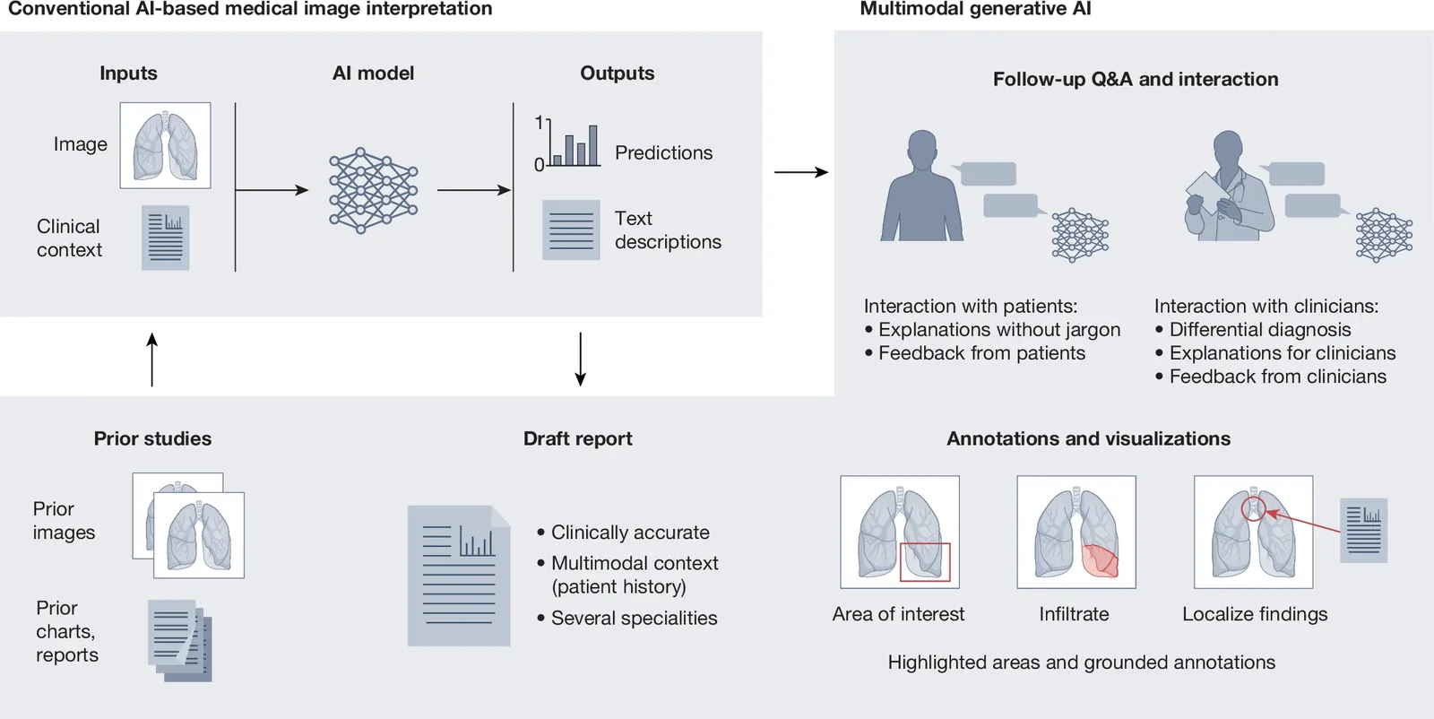
Multimodal generative AI for medical image interpretation
Radiology reports are written by hand, one image at a time, by specialists who are in short supply. This Perspective synthesizes the progress and open problems in generative models that draft reports directly from medical images, with attention to validating accuracy, exposing reasoning, and capturing the nuance of a real impression.
It proposes that such a system should enter practice the way a trainee does: supervised at first, calibrated to the local hospital, and given autonomy only as it earns it.
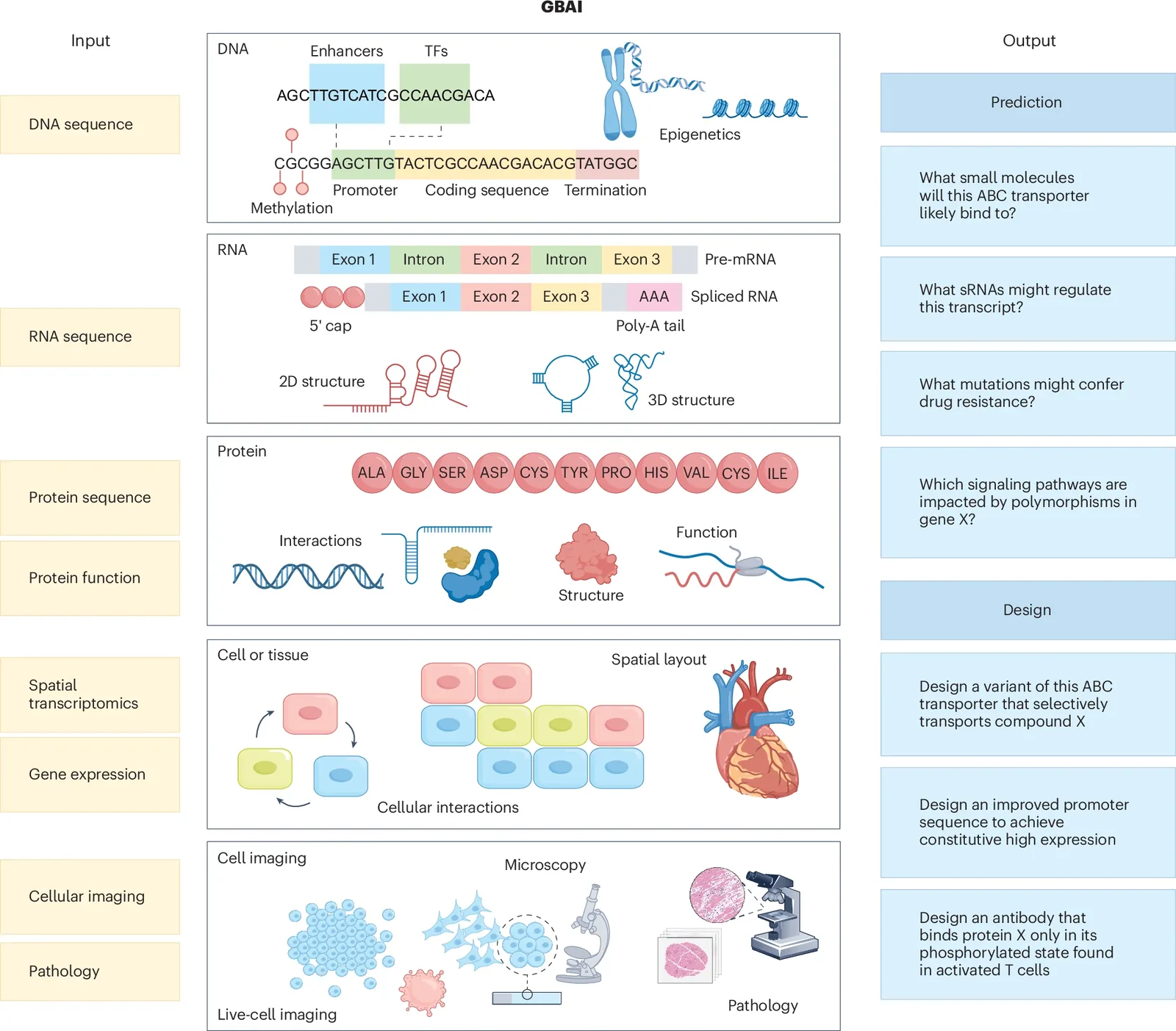
Generalist biological AI in modeling the language of life
Biology's AI models are specialists: one for protein structure, another for regulatory DNA, another for single-cell expression. This Review maps what those models have actually delivered across DNA, RNA, proteins, and cells, and makes the case for generalist biological AI, systems that read and generate across all of them at once.
It sets out what has to change in data, scale, and experimental validation for these models to become usable scientific instruments, from therapeutic design to virtual cells.
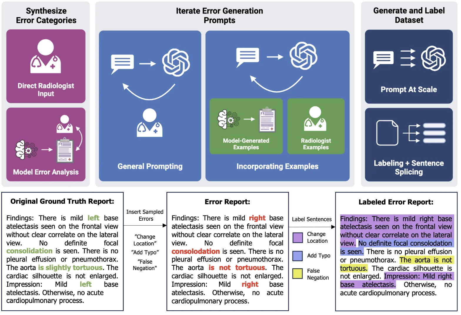
ReXErr: synthesizing clinically meaningful errors in radiology reports
You cannot test whether a report generation model makes dangerous mistakes without a labelled set of dangerous mistakes. ReXErr combines radiologist input with iterative language-model prompting to inject realistic errors, wrong laterality, false negations, missed findings, into real chest X-ray reports, each one labelled by type at the sentence level.
Released on PhysioNet as ReXErr-v1: over a million erroneous sentences derived from MIMIC-CXR, for evaluating and training report generation systems.
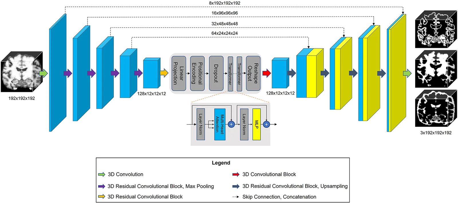
Improving across-dataset brain tissue segmentation using transformer
Brain tissue segmentation models trained on one scanner tend to fall apart on another. TABS is a hybrid 3D CNN–Transformer that segments grey matter, white matter, and CSF from T1-weighted MRI, validated across four multi-site datasets spanning vendors, field strengths, scan parameters, and neuropsychiatric conditions, plus test–retest scans.
It generalised better than every benchmark it was compared against, and the code is open.
Identifying bias in models that detect vocal fold paralysis from audio
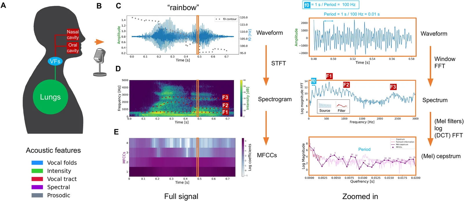
A voice recording could screen for vocal fold paralysis long before a patient reaches a laryngoscope. On 77 patients and 77 matched controls, our models reached an AUC of 0.87, above clinicians rating the same recordings, and we used SHAP to ask what the models were actually listening to.
Some of it was real. Some of it was artefact: differences in recording duration and intensity introduced by how the data had been stored, which a model could exploit without hearing anything clinical. We traced these biases and mitigated them.
Detecting schizophrenia with 3D structural brain MRI using deep learning
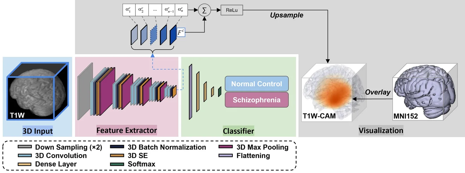
A 3D convolutional network that separates schizophrenia from healthy controls on a single conventional T1-weighted scan, evaluated across three open datasets, with an AUC of 0.987 on unseen MRI.
No psychiatrist will act on a black box. Alongside the classifier we used class activation maps to localise which regions each prediction depended on; subcortical structures and the ventricles dominated, consistent with the known neuroanatomy of the disease, so the output can be checked rather than trusted.
Adapt adult radiograph interpretation models to pediatric and neonatal care through continual learning across hospitals. Includes a pretrained model and a demo for ETT tip and carina localization.
Over a million clinically meaningful chest X-ray report errors derived from MIMIC-CXR, labelled by error type, released on PhysioNet for evaluating report generation models.
Transformer-based automated brain segmentation for T1-weighted MRI, built to hold up across datasets, vendors, and field strengths it wasn't trained on.